Andiprion paxtonae Hints, Tonarova et Eriksson, 2017
| ID | 15434 |
|---|---|
| Fossil group | Eunicida |
| Taxon | Andiprion paxtonae |
| Author | Hints, Tonarova et Eriksson, 2017 |
| Reference | Hints et al., 2017a |
| Parent taxon | Andiprion |
| FAD | Dapingian |
| LAD | Dapingian |
Type specimens
| Type | No | Type locality | Type horizon | Remarks | Reference | ||||
|---|---|---|---|---|---|---|---|---|---|
| holotype | IANIGLA, Instituto Argentino de Nivología, Glaciología y Ciencias Ambientales, Mendoza, Argentina | PI 3117 | Río Capillas section, Sierras Subandinas, northwestern Argentina | Dapingian | Transition between the Zanjón and Labrado formations, Dapingian, Middle Ordovician | Hints et al., 2017a |
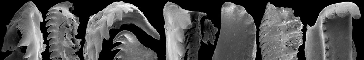
Images

Description(s)
Hints et al., 2017a:
Diagnosis. Labidognath-type apparatus consisting of basal plate, left and right MI, and anterior maxillae of which only left MII and MIII are known; carriers most likely present, probably subtriangular; precise size relationships of elements in apparatus unknown.
Description. Right MI: L=138–405 µm, L/W=1.7–2.7. Elongated jaw. Anterolateral margin from anterior towards posterior with convex and then concave part, ramus pointed, directed postero-laterally, extending to 0.25 to 0.4 of jaw length. Anterolateral part of jaw with fang and ramus loosely connected with rest of jaw, commonly showing suture line (Figs. 3E, 3G, 2P); if broken off, anterolateral margin is straight (Fig. 2F). Shank slender, posteriorly rounded. Inner margin straight or slightly concave in anterior third to half of jaw length, then turns sharply at 120–140˚ angle and runs towards posterior end of jaw. Dentary contains 12–18 denticles, which are often smaller and slenderer in the anterior third of dentary, the largest denticles occur around mid-length decreasing in size posteriorly. In ventral view myocoele opening gaping; cover missing.
Left MI: L=166–430 µm, L/W=2.4–4.0 (holotype: L=230 µm, W=81 µm, L/W=2.8). Outer margin runs posterolaterally almost straight or with convex and concave part, then turns and runs straight to meet posterior margin; outer face has maximum width around 0.35–0.5 of jaw length. Posterior margin straight, directed anterolaterally, corresponds to 0.5–0.8 of jaw width. Inner wing subrectangular, ca 0.3 of jaw width, occupying 0.4–0.6 of jaw length. Dentary has 13–17 denticles, which are slender in the anterior third and largest and sturdiest around mid-length decreasing in size posteriorly. In ventral view myocoele opening gaping; cover missing.
Basal plate: L=166 µm, W=86 µm, L/W=1.9. Subtriangular jaw, widest at mid-length; posterior margin straight, directed antero-laterally, corresponding to one third of jaw width; dentary with 20 tightly packed denticles directed laterally, decreasing in size posteriorly; posterior quarter undenticulated.
Left MII: L=190 µm. Slender jaw with pointed ramus directed posterolaterally and extending to nearly half of jaw length. Similarly to right MI, ramus with first denticle may separate from rest of jaw along suture line (Fig. 3D). Dentary with 12 denticles of which two anterior ones are more slender, largest denticles occur around mid-length, decreasing in size posteriorly, similarly to left and right MI. Inner wing subtriangular, widest at around mid-length.
Left MIII: L=180 µm. Similar to left MII, but relatively shorter; inner wing occupies ca half of jaw length; dentary with 10 posteriorly decreasing denticles not differentiated in shape.
Carriers: Unknown, but the posterior margin of the left and right MI indicates that carriers with a wide anterior margin were present in the apparatus. Probably they were subtriangular in shape, like those recorded in Kadriorgaspis.
Variability. Assessing the variability of A. paxtonae is complicated as most of the jaws to hand are partly flattened or otherwise deformed and broken. For instance, the length/width ratio and element size therefore vary greatly. However, the type of denticulation and the main outline, especially the shape of the inner and outer wings in both MI, are rather constant. One small left MI (Figs. 2K, 3A) is slightly different from the other specimens in having a posterior margin directed more perpendicular to the dentary and an inner wing which is longer and anteriorly wider. These minor differences are, however, regarded to fall within the intraspecific variability.
Comparison. The individual jaws of A. paxtonae show resemblance to those of several different taxa, but the entire apparatus has no closely matching forms. An unpublished Dapingian collection from northern Estonia contains a single left MI (illustrated for comparison in Fig. 2U), which is notably similar to that of A. paxtonae, especially the element shown in Figs. 2K and 3A. The similar morphology and closely matching age suggest that these two taxa may be congeneric, but further comparison awaits additional material from the Baltic region. The left MI of A. paxtonae shows also similarity to the corresponding jaw of polychaeturids in possessing a wide and straight posterior margin, such as in Pteropelta kielanae and P. huberti (see Hints 1998; Hints & Eriksson 2010), and some ramphoprionids, like species of Protarabellites (see e.g., Hints 1998; Eriksson 2001), but other jaws are different. The right MI shares the long shank and narrow ramus with certain polychaetaspids. The basal plate of A. paxtonae resembles that of Conjungaspis minutus Hints, 1999 in having similar denticulation, but differs in the posterior termination. The left MI, and to some extent also the basal plate, of A. paxtonae also resemble those elements of Kadriorgaspis Hints & Nõlvak, 2006.
Remarks. The jaw apparatus of Andiprion is reconstructed from isolated jaws using both morphological criteria and relative abundance of individual jaws. For instance, the size range, type of denticulation and shape of the anterolateral margin in both left and right MI suggest that these jaws belong to one and the same apparatus and hence a single, multi-element-based species. This is supported by the nearly equal number of corresponding left and rightMI recorded in the sample and the lack of feasible alternative apparatus configurations. In addition to posterior maxillae the collection contains a basal plate and a left MII. The basal plate is compatible with the same apparatus architecture as its shape fits well into the bight of the right MI and those elements combined have a posterior margin similar to that of the left MI. Both the basal plate and left MI must fit with the posterior carriers; the plate-like, supporting elements that are symmetrical in all taxa hitherto known. The left MII shares the main outline and denticulation type with that of the right MI, and has also a similarly attached ramus.